CYTONOTE 1W

Time-lapse images of cells and real time analysis from inside your incubator
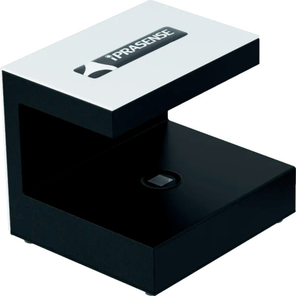
A small footprint device that stays always in focus with Petri dishes, T-flasks, slides or microfluidic chips
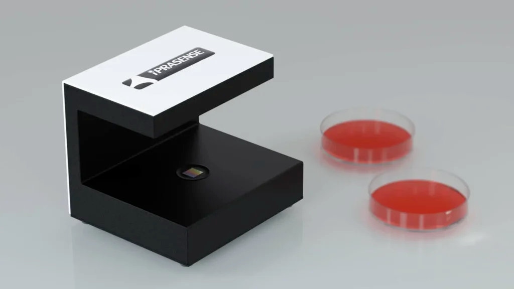
The CYTONOTE 1W uses our patented lens-less imaging technology in a tiny device to fit with any incubator. The CYTONOTE 1W is the simplest live cell-imaging system designed for recording cell movies and analyzing a variety of cell culture from the incubator. The innovative and patented “Lens-less imaging” technology pushes the boundaries of microscopy with its super wide field of view and its capability to capture and analyze precisely several thousands of cells without any focus and brightness settings. This technology simplifies the Live Cell Imaging technique and transforms the complex and expensive microscope method into a cost-effective solution.
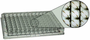
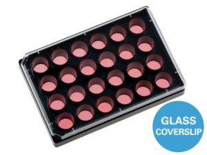
The CYTONOTE 1W is designed to be compatible with every standard lab dishes, T-flask or slides. Select your container and place it on the CYTONOTE 1W. The acquisition time between two measurements is 20 minutes by default, but you can change this time if you wish. By performing acquisitions over a give period of time you will be able to obtain time lapses and movies of your cells.
As explained, no need to adjust focus, brightness or any other parameters, our algorithm will reconstruct cells wherever cells are located… from few micrometers to some millimeters! This characteristic makes the CYTONOTE 1W an extremely robust device not fearing temperature variation, cells descent inside Matrigel, or any other parameter which can affect traditional focus adjustment.
The CYTONOTE 1W works on an ultra-wide field of view, where all data recovered from picture represent your cell culture.
The image analysis and the result from the CYTONOTE 1W are performed from the HORUS dedicated Software. HORUS is application oriented: it provides quantitative confluence determination, cell size, cell tracking… Full field image (30mm2) of the sample is stored and can be accessed and zoomed at any time.
Features of our Live Cell Imaging System
- Tiny device
- Directly from your incubator
- Time-Lapse & Movie
- Many cell culture applications
- Robust
- High Contrast
- Always in focus
- Wide field of view
Applications of our Live Cell Imaging System
- Real-Time Imaging of your cell culture
- Time-Lapses and Movies of your cells
- Confluence analysis
- Growth curves
- Migration test
- Proliferation test
- Cell morphology
- Cell population
- Cell attachment/detachment
- Cell tracking
Accessories & Consumables of our Live Cell Imaging System
- HORUS Software
- 21 CFR Part 11
- Scratch Assay Analysis module for HORUS
- Cell Tracking Analysis module for HORUS
- Cell Tube Formation Analysis module for HORUS
- CYTONOTE Cable (2 meters)
- Laptop Computer
- IQ/OQ
| Cells | Eucaryotic cells : Adherent monolayer, Suspension cell at bottom of culture ware or in micro-slides, 3D spheroids |
| Media | Liquid or Semi-solid (collagen) |
| Culture vessels | Standard plastic petri dish, culture flask, multiwell plate, max height 55mm |
| Resolution | 1 micron |
| Field of view | 30 mm² (6,5 mm x 6,5 mm) |
| Working distance | 0 to 5 mm |
| Image rate | 1,5 image/min |
| Light source | LED |
| Sensor | CMOS 10 Mpix |
| Image format |
|
| Enclosure | Stainless steel |
| Size | 12 * 11 * 10 cm |
| Weight | 1 kg |
| Power supply | USB |
| Pharmaceutical industries |
|