µ-Plate 384 Well

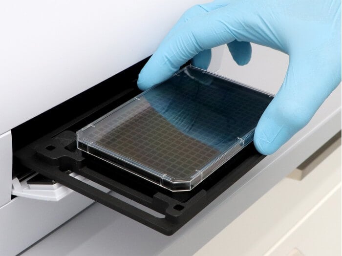
- Microtiter plate compatible with robotics, due to an ANSI/SLAS (SBS) standard format
- Brilliant cell imaging due to the excellent inner well and whole plate flatness
- Black walls provide low well-to-well crosstalk for fluorescence microscopy
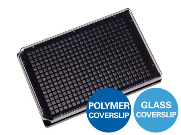
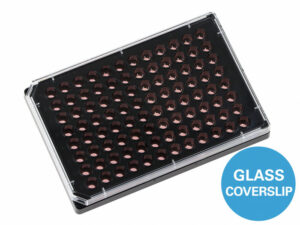
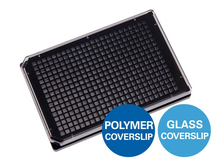
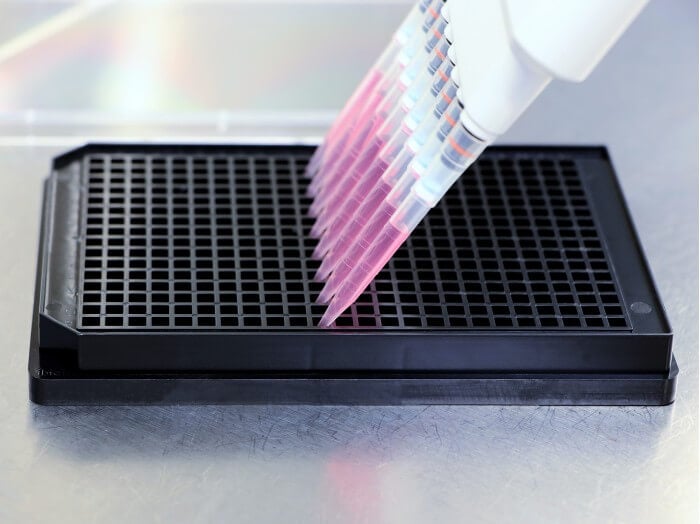
A black 384 well plate with square wells for high-throughput screenings
Applications
Cell Cultivation and Imaging
- Cultivation and high-resolution microscopy of cells
- Parallel cell culture experiments with minimal cell numbers and reagent volumes
- High-sensitivity immunofluorescence stainings and protein localization studies
- Live cell imaging and time-lapse studies over extended time periods
- Large-scale gene and protein expression analysis, transfection assays
- Drug discovery studies, cytotoxicity assays, and toxicology screenings with clear visibility of cell morphology
Microscopy Techniques
- Brightfield and widefield fluorescence microscopy for cell monitoring on inverted microscopes
- Automated, multiparametric high-content screening (HCS) and high-throughput screening (HTS) with plate readers
- Super-resolution microscopy (STED, (F)PALM, (d)STORM, SIM) and TIRF (best output with #1.5H Glass Coverslip)
- Confocal microscopy, two-photon microscopy, FRAP, FRET, FLIM, and LSFM
Technical Features
Precision, Quality, and Biocompatibility
- µ-Plate (microtiter plate) in ANSI/SLAS (SBS) format
- Ready-to-use for lab automation and high-throughput with robotics, fluorescence scanners, and plate readers
- Twelve round wells with standard numbering (letters A–P and numbers 1–24)
- Excellent inner well flatness and whole plate flatness
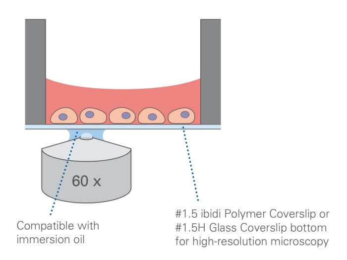
- #1.5 ibidi Polymer Coverslip or #1.5H Glass Coverslip bottom with excellent optical quality
- No autofluorescence due to black walls
- Compatible with staining and fixation solutions
- Compatible with immersion oil
- Made from non-cytotoxic, biocompatible materials
- Sterile with single packaging
Product Versions
- With #1.5 ibidi Polymer Coverslip (different surfaces available) or #1.5H Glass Coverslip Bottom
How Does the µ-Plate 384 Well Work?
What Is the Principle of the µ-Plate 384 Well?
The µ-Plate 384 Well consists of 384 independent wells with a thin #1.5 ibidi Polymer Coverslip or #1.5H Glass Coverslip bottom. The ibiTreat (tissue culture-treated) surface has been designed for optimal cell adhesion on the #1.5 ibidi Polymer Coverslip bottom. The rigid glass bottom provides excellent flatness for consistent focus across the plate, supporting high-resolution microscopy and screening workflows.
What Is the µ-Plate 384 Well Used for?
The µ-Plate 384 Well is designed for high-content and high-throughput imaging applications, including immunofluorescence staining, compound screening, transfection assays, and live-cell imaging. The glass bottom is particularly suitable for TIRF, single-molecule imaging, and super-resolution microscopy where tight thickness tolerances and high optical performance are required.
Is the µ-Plate 384 Well Compatible With Automated Systems?
Yes. The plate follows the ANSI/SLAS (SBS) footprint and is compatible with robotic liquid handling, automated microscopy platforms, fluorescence scanners, and plate readers, enabling reproducible positioning and streamlined screening workflows.
Is the µ-Plate 384 Well Suitable for Live Cell Imaging?
Yes. The coverslip-like bottom provides the optical performance required for long-term live cell imaging. For stable temperature, CO2, and humidity, use it together with a Stage Top Incubator.
Do I Need to Coat the Glass Bottom for Cell Adhesion?
Depending on your cell type, coating may be required to promote attachment on the glass surface. Common coatings include extracellular matrix proteins (e.g., collagen, fibronectin, laminin) or poly-D-lysine for sensitive cell types. Choose the coating based on the biological model and assay requirements.
How Do I Conduct Immunofluorescence Stainings With the µ-Plate 384 Well?
The µ-Plate 384 Well is well suited for immunofluorescence (IF) assays. Separate wells enable multiple conditions and replicates in one plate, with convenient washing and staining directly in the wells. The integrated #1.5 or #1.5H imaging bottom provides excellent optics for widefield fluorescence and confocal microscopy, no additional coverslip mounting or sample transfer is required.
| Outer dimensions (l × w) | 127.7 × 85.5 mm |
| Number of wells | 384 |
| Height with/without lid | 17.2 / 15.0 mm |
| Well-to-well distance | 4.5 mm |
| Single well dimensions (l × w) | 3.4 × 3.4 mm |
| Single well depth | 12.7 mm (88401, 88406) 12.8 mm (88407) |
| Volume per well | 50 µl |
| Growth area per well | 0.11 cm² |
| Coating area per well using 50 µl | 0.8 cm² |
| Bottom material | #1.5 ibidi Polymer Coverslip (different surfaces available) #1.5H Glass Coverslip (uncoated and untreated) |